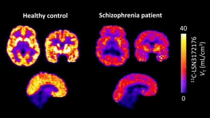
Summary: Researchers delivered the world’s first in vivo (in a living organism) evidence of widespread muscarinic acetylcholine M1 receptor deficits in schizophrenia. Utilizing a pioneering, highly selective PET radiotracer, the researchers discovered that living schizophrenia patients exhibit a striking 13% to 19% reduction in M1 receptor availability across multiple core cognitive regions of the brain. The discovery validates a major shift toward non-dopaminergic precision treatments.
Key Facts
- The M1 Muscarinic Deficit Discovered: The high-resolution PET imaging revealed that patients diagnosed with schizophrenia experience a significant 13% to 19% drop in functional M1 receptor availability across widespread cortical and subcortical regions compared to completely healthy control peers.
- Direct Alignment with Cognitive Deficits: Crucially, the scarcity of M1 receptors was much more strongly linked to objective measures of cognitive impairment than to the severity of positive psychotic symptoms. This reveals that M1 dysfunction is a primary biological driver behind the memory, learning, and executive processing challenges that heavily disrupt daily life.
- Bypassing the Postmortem Limitation: For decades, neurobiology was trapped because nearly all evidence connecting acetylcholine to schizophrenia came from postmortem tissue samples. This left it entirely unclear if the deficits were present during life or simply caused by decades of heavy medication use. This study provides the definitive in vivo baseline confirmation.
- Validating Next-Gen Non-Dopaminergic Drugs: The study arrives at an extraordinary historical turning point in psychiatry. The findings provide immediate biological reinforcement for newly approved therapies like xanomeline–trospium (COBENFY™), the first antipsychotic medication approved in over 70 years that treats schizophrenia through a non-dopaminergic, muscarinic mechanism.
- Strengthening Precision Psychiatry Pathways: While this trial did not actively track medication responses, Dr. Rajiv Radhakrishnan notes that M1 receptor PET scans could eventually be used as a precision diagnostic tool to identify distinct biological subgroups of patients, allowing doctors to match individuals to targeted treatments.
- A New Focus on G-Protein-Coupled Receptors: The data cements M1 receptors, which are widely distributed G-protein-coupled receptors crucial for synaptic plasticity, as an essential target for modern psychiatric drug development and diagnostic focus.
Source: Elsevier
A groundbreaking study using positron emission tomography (PET) imaging has found that patients with schizophrenia had significantly lower muscarinic acetylcholine M1 receptor availability (~13% to 19%) across multiple brain regions compared with healthy individuals. Reductions in M1 receptors affect several brain regions involved in cognition, learning, memory, and executive function.
The findings in Biological Psychiatry, published by Elsevier, provide the first in vivo evidence supporting widespread M1 receptor deficits in schizophrenia.
Schizophrenia is a serious mental disorder that is heterogeneous in its expression and biology. For many years, abnormalities in the brain’s muscarinic acetylcholine system, particularly the M1 receptor, have been implicated in the pathophysiology of schizophrenia. However, nearly all of the evidence came from postmortem studies, making it impossible to determine whether these abnormalities were present in living patients or how they related to clinical symptoms.
“The development of a novel PET radiotracer for the M1 receptor provided a unique opportunity to directly measure M1 receptor availability in the living brain,” explains co-lead investigator Deepak C. D’Souza, MBBS, MD, Department of Psychiatry, Yale University School of Medicine; Psychiatry Service, VA Connecticut Healthcare System; and Abraham Ribicoff Research Facilities, Connecticut Mental Health Center.
“Although receptor availability is not identical to receptor density, it is widely accepted as a useful proxy for the brain’s functional M1 receptor system. This allowed us, for the first time, to confirm that muscarinic dysfunction is a feature of schizophrenia in living patients.”
“The study’s findings were robust across multiple methods of PET quantification and remained significant after accounting for potential confounding factors, including gray matter differences and partial-volume effects” says co-first author Tommaso Volpi, MD, PhD, Associate Research Scientist in Radiology and Biomedical Imaging, Yale University School of Medicine.
Researchers highlighted that M1 receptor availability was more strongly associated with measures of cognition than with the severity of psychotic symptoms, suggesting that M1 dysfunction may be particularly relevant to the cognitive impairments that are among the most disabling aspects of schizophrenia.
The pharmacological treatment of schizophrenia has been dominated by dopamine D2 receptor antagonist/agonists commonly referred to as antipsychotics. Their limited efficacy, especially for negative and cognitive symptoms, and their significant side effects have spurred the search for drugs with alternative mechanisms of action. M1 receptors are G-protein-coupled receptors that are present throughout the cortex and subcortical regions. They are now considered an important focus of the underlying neurobiology and treatment of schizophrenia.
John Krystal, MD, Editor of Biological Psychiatry, comments, “This study is particularly interesting in light of the emergence of M1 and M4 muscarinic agonist drugs as pharmacotherapies in schizophrenia. These findings are particularly timely given the recent approval of xanomeline–trospium (COBENFY™), the first antipsychotic medication in more than 70 years to treat schizophrenia through a primarily non-dopaminergic mechanism of action.”
“Although our study did not evaluate treatment response, it strengthens the biological rationale for developing muscarinic-based therapies and raises the possibility that M1 receptor imaging could eventually help identify biologically distinct subgroups of patients and inform future precision medicine approaches,” concludes co-lead investigator Rajiv Radhakrishnan, MBBS, MD, Department of Radiology and Biomedical Engineering and Department of Psychiatry, Yale University School of Medicine; Psychiatry Service, VA Connecticut Healthcare System; and Abraham Ribicoff Research Facilities, Connecticut Mental Health Center.
Key Questions Answered:
A: The barrier wasn’t a lack of interest, but a lack of specialized technology. In neuroimaging, to see a specific receptor on a brain scan, you need a “radiotracers”, a specially engineered molecule injected into the blood that can cross the blood-brain barrier, cleanly bind to only one specific type of receptor, and emit a faint signal that a PET scanner can pick up. For decades, scientists could only manufacture radiotracers for dopamine or serotonin systems. Muscarinic acetylcholine receptors were notoriously difficult to isolate. The development of this novel, highly selective M1 radiotracer by the Yale team finally gave medicine the precise key needed to unlock and view this system in a living human mind.
A: This is a vital technical distinction in molecular imaging. Receptor density refers to the absolute physical count of receptors built onto the surface of a cell, a metric that can typically only be counted under a microscope in postmortem tissue samples. Receptor availability, which is measured by a PET scan, tells us how many of those receptors are completely open, functional, and ready to bind with chemical signals in a living brain. While they are not perfectly identical, receptor availability serves as an outstandingly accurate proxy for measuring the real-time strength and health of the brain’s functional M1 network.
A: For more than 70 years, every single antipsychotic medication approved by the FDA relied on the exact same mechanism: slamming the brakes on the brain’s dopamine D2 receptors. While this can help quiet down acute hallucinations, it does very little to improve cognitive processing and often carries severe, exhausting side effects. The recent approval of xanomeline–trospium (COBENFY™) marked a historic shift because it completely ignores dopamine, operating instead as a muscarinic agonist to activate acetylcholine receptors. This Yale study provides the missing piece of baseline science that validates this drug’s entire design, proving that living patients suffer from a significant deficit in the exact M1 receptors this new class of medicine is built to rescue.
Editorial Notes:
- This article was edited by a Neuroscience News editor.
- Journal paper reviewed in full.
- Additional context added by our staff.
About this schizophrenia research news
Author: Eileen Leahy
Source: Elsevier
Contact: Eileen Leahy – Elsevier
Image: The image is credited to Biological Psychiatry / Volpi et al.
Original Research: Open access.
“Lower Muscarinic M1 Receptor Availability in Schizophrenia: In Vivo PET Evidence” byDavid Labaree, Deepak C. D’Souza, Mika Naganawa, Nabeel Nabulsi, Rachel Hird, Rajiv Radhakrishnan, Richard E. Carson, Soheila Najafzadeh, Swanee Jacutin-Porte, Tommaso Volpi, Yiyun Huang. Biological Psychiatry
DOI:10.1016/j.biopsych.2026.06.002
Abstract
Lower Muscarinic M1 Receptor Availability in Schizophrenia: In Vivo PET Evidence
Background
There is growing interest in the contributions of brain muscarinic M1 receptors to the neurobiology of schizophrenia (SZ). Postmortem evidence strongly indicates M1 deficits in SZ patients. Recently, the FDA approved COBENFYTM, which contains M1/M4 agonist xanomeline, as the first non-dopaminergic medication for SZ. The development of the PET ligand 11C-LSN3172176 enables in vivo quantification of brain M1 availability in SZ and its relationship to clinical features.
Methods
M1 availability in SZ patients (n=16) was compared to age/sex-matched healthy controls (HC) (n=16) using 11C-LSN3172176 and the High-Resolution Research Tomograph. Distribution volume ratio relative to the centrum semiovale (DVRCS) and distribution volume (VT) were measured. Regional variation in percent gray matter fraction (%GM) was included as a covariate to account for potential atrophy.
Results
Compared to HCs, patients with SZ showed significantly lower M1 availability across several cortical and subcortical regions, with large effect sizes: frontal (DVRCS: -13%), temporal (DVRCS: -15%; VT: -12%), parietal (DVRCS: -14%; VT: -11%), occipital (DVRCS: -16%; VT: -13%), caudate (DVRCS: -19%; VT: -15%), putamen (DVRCS: -19%; VT: -17%), hippocampus (DVRCS: -13%), amygdala (DVRCS: -19%; VT: -16%). A subgroup of patients (DVRCS: 44%; VT: 27%) showed larger whole-brain M1 deficits (>20% from the mean of HCs). Exploratory analyses suggested associations between M1 availability and selected clinical measures.
Conclusions
These in vivo findings together with postmortem data warrant further characterization of M1 deficits in SZ and highlight M1 as a therapeutic target and potential biomarker for muscarinic-based treatments.